
Here we report an unusual case of. Hes got Koplik spots on the buccal mucosa.
Hematoma
It is one of the.

. The swelling and pain usually reduces within one or two weeks depending on the size of hematoma. Lesions are erythematous and edematous. As salivary glands are involved.
There was no clear identifiable left Whartons duct. Epidur hemor w LOC w death due to oth causes bf consc init. Hematoma can occur in any organ of the body.
This means that the tumor has spread to lymph nodes in the neck. However in buccal cancers the primary cancer in the mouth is usually noticed before it reaches these lymph nodes. Buccal mucosa is a common site of thrombus presentation.
ICD-10-CM Diagnosis Code S064X8A convert to ICD-9-CM Epidural hemorrhage with loss of consciousness of any duration with death due to other causes prior to regaining consciousness initial encounter. Also noted was a small area of left lower lip buccal mucosa avulsion diffuse submental swelling and ecchymoses. However in buccal cancers the primary cancer in the mouth is usually noticed before it reaches these lymph nodes.
The full SNOMED CT. A bare metal stent was implanted in the LAD. S005 Powered by X-Lab.
Also noted was a small area of left lower lip buccal mucosa avulsion diffuse submental swelling and ecchymoses. Abstract and Figures Our previous studies based on intraoral dissection of fresh cadavers revealed that the fissure and loose connective tissues. Additional Symptoms Loose teeth or dentures that dont fit correctly.
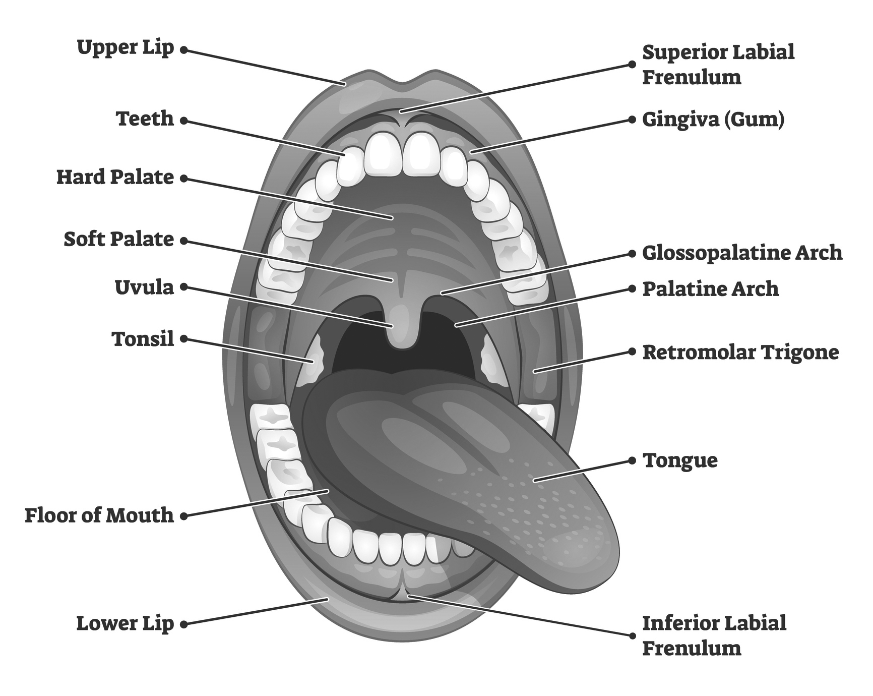
The inner lining of the cheeks and lips which is an anatomic region that includes all the mucous membrane lining of the inner surface of the cheeks and lips from the line of contact of the opposing lips to the line of attachment of mucosa to the alveolar ridges and pterygomandibular raphe which occupies an area of 5060 cm2. The oral mucosa has a thick elastin-rich epithelium which is resistant to shearing or stretching forces. In the following days ulcerative erosions with whitish-yellow exuadate appear.
Benign proliferation of endothelial lining of blood vessels may clinically present as a raised bluish hemangioma of the oral mucosa. The lower lip and tongue were diffusely enlarged with the tongue being displaced posterosuperiorly obstructing the oral cavity and oropharynx and protruding from the mouth. A hematoma is collection of blood which has leaked out from the blood vessel into the surrounding tissue space.
Oral hematoma can occur due to traumatic injury in or outside of the mouth. Lipoma accounts for 1-4 of benign neoplasms of mouth affecting predominantly the buccal mucosa floor of mouth and tongue. Epidural hematoma with loss of consciousness and death.
Mouth cancers can arise in this area and are often associated with previous lichen planus or sometimes when patients have been previous tobacco or pan chewers. Hemangiomas are soft dark red to blue sessile or pedunculated lesions with smooth or lobulated surfaces that blanch on compression. Postoperative complications were not associated with age or sex but were associated with recipient site and tumor stage.
Hematomas were 188 95 CI 16-217 times more common at the buccal mucosa. Translations in context of buccal mucosa in English-Spanish from Reverso Context. Lipomas when superficially placed show yellowish surface discoloration and hemangiomas usually have reddish blue to deep blue color.
In some cases the first sign of buccal cancer could be a lump in the neck. Properties of Oral Mucosa. Buccal and labial mucosa have non-keratinized stratified squamous avascular epithelium with vascular connective tissue called lamina propria.
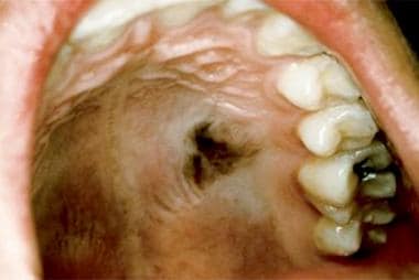
Generally buccal mucosa is affected by radiation treatment of head and neck tumors. In the second day of the admission there was a 3020 mm hematoma in the buccal mucosa of the patient Figure 1. These cancers have sometimes unfortunately been under-treated but with modern free flap reconstructions good surgical clearance can be.
- Hematoma of buccal mucosa - Haematoma of buccal mucosa - Hematoma of buccal mucosa disorder Hide descriptions. There are connective tissue projections from the lamina propria into the epithelium which. Buccal MucosaThe buccal mucosa is the mouth lining to the cheek.
Hemangiomas occur mostly on the lips buccal mucosa tongue and palate. Overall complications were 03 95 confidence interval CI 01-11 times less common at the buccal mucosa than at other recipient sites. The patient was clinically stable.
They present by the first month of. This tool allows you to search SNOMED CT and is designed for educational use only. When a pocket of blood forms in the extra-vascular space of oral cavity the condition is called oral hematoma.
After radiotheraphy at the end of first week the first oral manifestations can appear.

Intra Oral Photograph Showing Oral Hematoma On The Right Buccal Mucosa Download Scientific Diagram
Disorders Of Oral Pigmentation Background Pathophysiology Etiology
How Long Does It Take For An Oral

Pathology Injurie Of Soft Tissues Flashcards Quizlet
Injection
Trismus

Intraoral Photograph Showing A Pouch On The Right Buccal Mucosa Download Scientific Diagram

Figure 5 Oral Lesions And Lymphoproliferative Disorders

Intraoral Examination Normal Structures As Well As Landmarks Used For Placing Local Anesthetic Pocket Dentistry

Buccal Mucosa Alveolus Otorhinolaryngology Portal

Erythematous Oral Lesions When To Treat When To Leave Alone Consultant360

Pdf Melanoma De La Mucosa Oral Casos Clinicos Y Revision De La Literatura Semantic Scholar
Oral Path Mucosal Reactive Lesions Flashcards Quizlet

Oral Maxillofacial Regional Anesthesia Nysora Nysora
An Anatomical Review Of Trauma To The Mouth And Throat 2020 11 24
Extravascular Blood Lesions A Guide To Clinical Differential Diagnosis Of Oral Mucosal Lesions Continuing Education Course Dentalcare Com

Photographs Of The Right And Left Buccal Mucosa Of A Male Patient Download Scientific Diagram
2

Buccal Mucosa Alveolus Otorhinolaryngology Portal